| 目录号 | MX4021-100UL | 售价 | 996.00元 | ||||||||||||||||||||||||||||||||||||||||||||||||||||||||||||||||||||||
| 规格 | 100μl | 运输温度 | 冰袋运输 | ||||||||||||||||||||||||||||||||||||||||||||||||||||||||||||||||||||||
| 其他名称 | PKH26 Red Fluorescent Cell Linker Kit for General Cell Membrane Labeling | 保存温度 | 2-8℃避光保存 | ||||||||||||||||||||||||||||||||||||||||||||||||||||||||||||||||||||||
| CAS号 | N/A | 有效期 | 1年 | ||||||||||||||||||||||||||||||||||||||||||||||||||||||||||||||||||||||
| 应用 | 细胞增殖和示踪研究 | 订购数量 |
|
||||||||||||||||||||||||||||||||||||||||||||||||||||||||||||||||||||||
|
产品简介: PKH26 细胞连接试剂盒(用于常规细胞膜标记)
产品关键词: PKH26;Calcein AM钙黄绿素;PKH67;CFDA SE;In vivo cell tracking 体内细胞示踪;Sigma MINI26;Phanos Technologies;
产品信息
【好消息】:应客户的要求,我司对试剂盒内的Diluent C可单独供应,产品信息如下:
产品描述 PKH26细胞连接试剂盒(用于常规细胞膜标记)(PKH26 Cell Linker Kit for General Cell Membrane Labeling)是一款基于荧光探针PKH26,用于常规细胞膜标记的检测试剂盒,适用于体外细胞标记,体外细胞增殖以及长期的体内细胞跟踪研究等。 PKH26是一种专利的膜标记探针,结构上带有一长脂肪族尾巴,能稳定插入细胞膜脂质区域。PKH26荧光处于黄-橙色光谱区间(见图1),最大激发波长为551nm,最大发射波长是567nm,与罗丹明或PE检测系统兼容。也可能用标准荧光素(Fluorescein)激发滤片,但荧光强度可能会有些降低。PKH26具最长的体内半衰期,超过100天,非常适用于体内细胞示踪、细胞增殖研究和其他长期实验。
稀释液C(Diluent C)是本试剂盒配套提供的一种水溶性溶液,特别设计的一种标记媒介能维持细胞活力,同时最大化染料溶解性和标记过程中的染色效率。Diluent C对哺乳动物细胞来说是等渗的,不含去污剂或有机溶剂,也不含生理盐和缓冲剂。根据细胞类型,染料标记后膜的内在化程度,标记细胞可能呈现不同的状态,从明亮,到均匀点状或斑驳状。然而,PKH26荧光在生理范围内不依赖于pH,且每个细胞的荧光强度通常不受染料位置的影响。
图1. PKH26的激发和发射光谱图
我司(懋康生物)提供三种规格的PKH26细胞连接试剂盒(用于常规细胞膜标记),其中: Mini Kit(CAT#:MX4021-100UL)建议用于小量或初步研究。当使用2ml染色体积(含2μM 终浓度的PKH26),本试剂盒含有足量的染料用于检测25次细胞样本(2×10⁷个细胞/次),和足量的Diluent C用于5次细胞样本(2×10⁷个细胞/次)。用户需根据细胞类型和实验目的来优化确定最佳的染料浓度。 Midi Kit(CAT#:MX4021-200UL)适用于中量研究。当使用2ml染色体积(含2μM 终浓度的PKH26),本试剂盒含有足量的染料用于检测50次细胞样本(2×10⁷个细胞/次),和足量的Diluent C用于30次细胞样本(2×10⁷个细胞/次)。用户需根据细胞类型和实验目的来优化确定最佳的染料浓度。 Maxi Kit(CAT#:MX4021-500UL)适用于大量或体内研究。当使用2ml染色体积(含2μM 终浓度的PKH26),本试剂盒含有足量的染料用于检测125次细胞样本(2×10⁷个细胞/次),和足量的Diluent C用于30次细胞样本(2×10⁷个细胞/次)。用户需根据细胞类型和实验目的来优化确定最佳的染料浓度 产品包装
|
|||||||||||||||||||||||||||||||||||||||||||||||||||||||||||||||||||||||||
|
产品编号 |
产品名称 |
规格 |
|
500T |
||
|
MX3009-5MG |
CFDA, SE细胞增殖示踪荧光探针 |
5mg |
|
500T |
||
|
MX3011-50UG |
Calcein, AM, Ultra Pure Grade钙黄绿素(绿色) |
50μg |
|
MX4021-100UL |
PKH26 Cell Linker Kit for General Cell Membrane Labeling PKH26细胞连接试剂盒(用于常规细胞膜标记) |
100μl |
|
1×50μg |
||
|
10mg |
||
|
10mg |
— —Written/Edited by V. Shallan【版权归MKBio懋康所有】
上海懋康生物科技有限公司是一家涉足于生命科学和生物技术领域研究的试剂、仪器和实验室消耗品与实验服务工作,主要从事细胞生物学、植物学、分子生物学、免疫学、生物化学、蛋白组学。生物制药与诊断试剂研发生产等领域。 本公司秉承“以人为本,以诚为信、合同守信”的经营理念。坚持"品质保障"的原则为广大客户提供优质产品。
延伸阅读(懋康生物独家整理)
鉴于近期大量的用户咨询PKH26用于外泌体染色(exosome stain)的方法,我司查阅相关文献资料,提供染色相关的一些信息,仅供交流学习用。
摘自文献1)Pužar Dominkuš P et al. PKH26 labeling of extracellular vesicles: Characterization and cellular internalization of contaminating PKH26 nanoparticles. Biochim Biophys Acta Biomembr. 2018 Jun;1860(6):1350-1361. PMID: 29551275
◇◇ 外泌体染色(Exosome staining)(简单流程见Fig 1)
外泌体冻干标准用超纯水重悬制备成1.0μg/μL溶液,之后用PKH26红色荧光检测试剂盒来标记。染色前,溶于Diluent C的PKH26在超纯水水浴锅于37℃孵育15min。对于对照样品,用不含颗粒的DPBS来替代外泌体标准。
① 染色流程A
PKH26用100μL diluent C稀释使其终浓度为8μM(染料浓度)。之后10μg外泌体(in 20μL DPBS,MKBio)用80μL diluent C稀释,加入染料溶液内,孵育5min,同时用枪轻轻混匀。用100μL不含外泌体的10% FBS (in DMEM,MKBio)来结合多余的染料。之后,外泌体用1ml DPBS稀释,4°C超速(100000×g)离心1h 10min。之后沉淀用50μL DPBS轻轻重悬。
② 染色流程B
外泌体的染色方法同流程A。之后稀释到2ml DPBS,转移到Vivaspin20 300-kDa MWCO filters,于4°C(4000×g)离心3min,之后用2ml DPBS清洗3次,最终用沉淀用50μL DPBS轻轻重悬。
③ 染色流程C
外泌体的染色方法同流程A。之后稀释到10ml DPBS,置于2ml 20%蔗糖(in DPBS,,MKBio)的上层。外泌体用4°C超速(100000×g)离心1h 10min。之后沉淀用50μL DPBS轻轻重悬。
④ 染色流程D
外泌体的染色方法同流程A。之后稀释到400μL DPBS,置于20%~60%非连续蔗糖梯度(in DPBS)的上层。外泌体用4°C超速(100000×g,MKBio)离心18h。分层3-6(密度范围1.08–1.15g/mL)吸在一起,分层7-10(密度范围1.17–1.23g/mL)吸在一起,每一份都稀释到30ml DPBS。外泌体用4°C超速(100000×g)离心1h 10min。之后沉淀用50μL DPBS轻轻重悬。
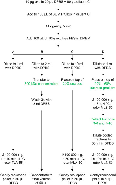
Fig. 1. Schematic representation of staining procedures used to label exosomes with PKH26 dye. Exosomes (exo) were labeled using staining procedures based on ultracentrifugation (procedure A), filtration through Vivaspin concentrators (procedure B), and sucrose cushion (procedure C) and sucrose gradient (procedure D) purification. Key differences between the procedures are highlighted by the green text.
◇◇ 外泌体检测(Confocal microscopy)
取小体积(6μL)轻轻混匀的PKH26标记对照和外泌体样本分别转移到包被有10%多聚L-赖氨酸的盖玻片上,然后封固在载玻片上,然后转移到共聚焦显微镜下观察。用561nm diode-pumped solid state laser line观察,用565nm-605nm宽带滤片来检测发射光。
◇◇ 外泌体染色结果分析
针对几种染色流程,发现①超速离心为基础的外泌体染色会产生大量的PKH26纳米粒子(nanoparticles),直径类似于PKH26标记的外泌体。②基于过滤(filtration)为基础的外泌体染色法典型的特征是PKH26标记颗粒的回收率低。③通过蔗糖缓冲层纯化的PKH26标记纳米粒子和PKH26标记外泌体根据大小能区分开。④PKH26标记纳米粒子和PKH26标记外泌体通过进入浓缩蔗糖梯度层能分开。
摘自资料2)来自网络资源。
PKH染料(常用的有PKH26-MX4021和PKH67)大量文献用来标记外泌体和胞外囊泡进行体外和体内示踪实验。以下步骤可参考用于外泌体标记以进行微囊泡示踪实验。
外泌体标记步骤:
(1)利用超速或微过滤的方式制备新鲜的外泌体沉淀。
(2)超速离心机冷却到2-8℃。将每个样本的多管离心沉淀聚合在一起,之后测定总体积。
(3)用Diluent C, MX4022将所有体积的沉淀稀释到高达1ml。
(4)确定最大体积的沉淀样本,加等体积的不含外泌体的培养基到一个新管内,用Diluent C稀释到1ml。
(5)加6μLPKH26到1ml Diluent C管内(步骤3和步骤4)。
(6)用枪连续性轻轻混匀30s。室温静置5min。
(7)加入含2ml 10% BSA(in PBS)来淬灭反应。用无血清培养基定容体积到8.5ml。
(8)制备0.971M的蔗糖溶液。用枪缓慢吸取1.5ml蔗糖溶液,加到管子底部,确保不会产生震荡。外泌体-PKH26 MX4021标记溶液加在蔗糖缓冲层(sucrose cushion)的上方。
(9)于2-8℃超速(190,000 G)离心2h。「注意:外泌体在沉淀内,绝大部分多余的染料在中间层」。小心吸取培养基和中间层。
(10)轻轻用枪将外泌体沉淀重悬在1X PBS。
(11)转移到Amicon 10kDa MWCO超滤管内。加9ml PBS,0.75ml培养基。
(12)于3000 x g离心40min,使得最终保留体积在0.5-1mL。
(13)从Amicon超滤管内吸取浓缩液,保存在冰上。尽快于合适的仪器下分析荧光信号。
引用文献:
[1]Yu Mengyu et al. Targeted exosome‐encapsulated erastin induced ferroptosis in triple negative breast cancer cells. Cancer Science. 2019;110:3173–3182.
(标记对象:外泌体)
Targeted exosome‐encapsulated erastin induced ferroptosis in triple negative breast cancer cells.
To quantify the amount of erastin@FA‐exo and erastin@exo taken up by MDA‐MB‐231 cells, lipophilic fluorescent dye PKH26 (MaoKang Biotechnology) was used to stain the exosomes. To detect the effect of FA receptor binding on cell uptake, culture medium containing 1.1 mg/mL of free FA was added to MDA‐MB‐231 cells to competitively inhibit FA receptors. After incubation for 6 hours, the cells were washed with PBS 3 times.Then erastin@FA‐exo was added and the cells’ uptake of the drug was observed.

[2]Yanlin Wang et al. Microfluidic Raman biochip detection of exosomes: a promising tool for prostate cancer diagnosis. Lab Chip, 2020,20, 4632-4637 https://doi.org/10.1039/D0LC00677G
(标记对象:外泌体)
PKH26 was obtained from Maokangbio Co., Ltd (Shanghai, China).
[3 ]Wu B, Ye Y, Xie S, Li Y, Sun X, Lv M, Yang L, Cui N, Che5n Q, Jensen LD, Cui D, Huang G, Zuo J, Zhang S, Liu W, Yang Y. Megakaryocytes Mediate Hyperglycemia-Induced Tumor Metastasis. Cancer Res. 2021 Nov 1;81(21):5506-5522. doi: 10.1158/0008-5472.CAN-21-1180. Epub 2021 Sep 17. PMID: 34535458.
(标记对象:血小板)
For the platelets adhesion assay, the platelet cell membrane was prelabeled by a PKH26 Cell Linker Kit (catalog no. MX4021, Maokangbio) for visualization.
[4] Che J, Wang H, Dong J, Wu Y, Zhang H, Fu L, Zhang J. Human umbilical cord mesenchymal stem cell-derived exosomes attenuate neuroinflammation and oxidative stress through the NRF2/NF-κB/NLRP3 pathway. CNS Neurosci Ther. 2024 Mar;30(3):e14454. doi: 10.1111/cns.14454. Epub 2023 Sep 12. PMID: 37697971; PMCID: PMC10916441.
(标记对象:外泌体)
The red fluorescent dye PKH26 (MaoKang Biotechnology, Shanghai, China) was used to label exosomes according to the manufacturer's instructions. The labeled exosomes were co-cultured with BV-2 cells or primary microglial cultures at a concentration of 10 μg/mL for 6 h, 12 h, and 24 h. Subsequently, the cells were fixed with 4% paraformaldehyde for 40 min and blocked using 5% bovine serum albumin for 1 h at room temperature. The cytoskeleton was stained with Actin-Tracker Green and the nucleus with DAPI. A confocal laser-scanning microscope (Leica TCS SP 5, Leica Microsystems GmbH) was used for imaging.

[5] Nie S, Zhang Z, Ji Y, Ding Q, Gong J, Xiao F, Chen L, Tian D, Liu M, Luo Z. CRIg+ macrophages deficiency enhanced inflammation damage in IBD due to gut extracellular vesicles containing microbial DNA. Gut Microbes. 2024 Jan-Dec;16(1):2379633. doi: 10.1080/19490976.2024.2379633. Epub 2024 Jul 18. PMID: 39024479; PMCID: PMC11259065.
(标记对象:细胞外囊泡 extracellular vesicles (mEVs))
To track mEVs entering cells and tissues, mEVs were labeled with PKH26 fluorescent dye using the PKH26 Cell Linker Kit (Maokang Biotechnology, Shanghai, China) according to the manufacturer’s instructions.
[6]Zong, Hf., Li, X., Han, L. et al. A novel bispecific antibody drug conjugate targeting HER2 and HER3 with potent therapeutic efficacy against breast cancer. Acta Pharmacol Sin 45, 1727–1739 (2024). https://doi.org/10.1038/s41401-024-01279-8
(标记对象:MCF10A细胞)
Normal breast tissue MCF10A cells were stained with 2 μM PKH26 (MX4201, Shanghai Maokang Biotechnology, Shanghai, China) following the manufacturer’s protocol.
[7]Yao XW, Liu ZY, Ma NF, Jiang WK, Zhou Z, Chen B, Guan WG, Yan JJ, Yang M. Exosomes from Adipose-Derived Stem Cells Alleviate Dexamethasone-Induced Bone Loss by Regulating the Nrf2/HO-1 Axis. Oxid Med Cell Longev. 2023 Feb 1;2023:3602962. doi: 10.1155/2023/3602962. PMID: 36778207; PMCID: PMC9908349.
(标记对象:外泌体)
Purified ADSCs-Exos were labeled with PKH26 (MaoKang Biotechnology, China), as instructed by the manufacturer.
[8] Chen, C., Wang, F., Cheng, C. et al. Cancer-associated Fibroblasts-derived Exosomes with HOXD11 Overexpression Promote Ovarian Cancer Cell Angiogenesis Via FN1. Reprod. Sci. (2024). https://doi.org/10.1007/s43032-024-01716-3
(标记对象:外泌体)
CAFs-Exo were labeled with PKH26 red fluorescent cell dye (MX4021, Shanghai Maokang Biotechnology Co., Ltd., Shanghai, China)
[9]Zhang B, Yang Y, Tao R, Yao C, Zhou Z, Zhang Y. Exosomes loaded with miR-665 inhibit the progression of osteosarcoma in vivo and in vitro. Am J Transl Res. 2022 Oct 15;14(10):7012-7026. PMID: 36398229; PMCID: PMC9641455.
(标记对象:外泌体)
The exosome was labeled by employing the PKH26 Cell Linker Kit (MX4021, Shanghai Maokang Biotechnology Co., Ltd., Shanghai, China) following the instructions.
[10]Zhang M, Yuan Q, Wang P, Zhang F, Wu D, Bai H, Liu J, Liu H, Yuan X. Bone Marrow Mesenchymal Stem Cell-Derived Nanovesicles Containing H8 Improve Hepatic Glucose and Lipid Metabolism and Exert Ameliorative Effects in Type 2 Diabetes. Int J Nanomedicine. 2024 Jul 3;19:6643-6658. doi: 10.2147/IJN.S455021. PMID: 38979532; PMCID: PMC11230129.
(标记对象:纳米囊泡 nanovesicles)
HepG2 cells were inoculated in confocal laser culture dishes with a density of 1×10⁶ cells per dish. After HepG2 cells were attached to the wall, BMSCs-NVs and H8-BMSCs-NVs solutions containing 10 μg/mL were put into 1.5mL EP tube, and PKH26 (2× 10⁻⁶m, Shanghai Maokang Biotechnology, MX4021) dye solution was added into the EP tube. After incubation at 37°C for 10 min, BMSCs-NVs and H8-BMSCs-NVs containing PKH26 dye solution27 were added to the confocal laser culture dish inoculated HepG2 cells for co-incubation. After 12 h, the confocal laser petri dish was taken out and washed 3 times with PBS (pre-heated at 37°C), and appropriate 4% paraformaldehyde solution was fixed for 10 min, then washed twice with PBS (30 s/ times), Triton100 (0.5%) for 2 min, and washed twice with PBS (30 s/ times). Phalloidin (Shanghai Maokang Biotechnology, MX4402) staining (in dark) was incubated at 37°C for 30min, washed with PBS twice (30 s/ time), DAPI staining for 2 min, and cleaned with PBS for 3 times. After that, the uptake of BMSCs-NVs and H8-BMSCs-NVs by HepG2 cells was observed under confocal laser microscope and images were collected.
[11]Jing Z, Zhang G, Cai Y, Liang J, Lv L, Dang X. Engineered extracellular vesicle-delivered TGF-β inhibitor for attenuating osteoarthritis by targeting subchondral bone. J Tissue Eng. 2024 Jul 24;15:20417314241257781. doi: 10.1177/20417314241257781. PMID: 39071897; PMCID: PMC11273819.
标记对象:(EVs)
PKH26 kit was purchased from Shanghai Maokang Biotechnology Co., Ltd.
[12] Sun J, Wei J, Zhang Y, Li J, Li J, Yan J, Guo M, Han J, Qiao H. Plasma Exosomes Transfer miR-885-3p Targeting the AKT/NFκB Signaling Pathway to Improve the Sensitivity of Intravenous Glucocorticoid Therapy Against Graves Ophthalmopathy. Front Immunol. 2022 Feb 21;13:819680. doi: 10.3389/fimmu.2022.819680. PMID: 35265076; PMCID: PMC8900193.
(标记对象:外泌体)
500 μg purified exosome suspension was taken and diluted to a volume of 500 μl with sterile 1× PBS. According to the instructions, exosomes were labeled with PKH26 staining kit (Maokangbio, Shanghai, China). After staining, the concentration of the stained exosome suspension was quantified by the BCA method, and then aseptic 1× PBS adjusted the exosomes to the appropriate concentration.
[13] Zhao X, Ge P, Lei S, Guo S, Zhou P, Zhao L, Qi Y, Wei X, Wu W, Wang N, Guo R, Yang N, Xiao Q, Zhang Q, Zhu H. An Exosome-Based Therapeutic Strategy Targeting Neuroinflammation in Alzheimer’s Disease with Berberine and Palmatine. Drug Des Devel Ther. 2023;17:2401-2420
https://doi.org/10.2147/DDDT.S417465
(标记对象:外泌体)
The PKH26 fluorescent probe was purchased from Maokangbio (Shanghai, China).
[14]Liu S, Fan M, Xu JX, Yang LJ, Qi CC, Xia QR, Ge JF. Exosomes derived from bone-marrow mesenchymal stem cells alleviate cognitive decline in AD-like mice by improving BDNF-related neuropathology. J Neuroinflammation. 2022 Feb 7;19(1):35. doi: 10.1186/s12974-022-02393-2. PMID: 35130907; PMCID: PMC8822863.
(标记对象:外泌体)
the PKH26 staining kit (MX4021-100UL, Maokangbio) was used to stain and label the exosomes
[15]Peng, H.; Du, F.; Wang, J.; Wu, Y.; Wei, Q.; Chen, A.; Duan, Y.; Shi, S.; Zhang, J.; Yu, S. Adipose-Derived Stem-Cell-Membrane-Coated PLGA-PEI Nanoparticles Promote Wound Healing via Efficient Delivery of miR-21. Pharmaceutics 2024, 16, 1113. https://doi.org/10.3390/pharmaceutics16091113
(标记对象:纳米载体)
PKH26 (MKbio, Shanghai, China; MX4021) was employed to label the membrane coatings according to the manufacturer’s instructions. PKH26-labeled nanocarriers were then incubated with different types of cells at 37 °C for 6 h. Cells were then fixed in 4% paraformaldehyde (Sigma-Aldrich, St. Louis, MO, USA; 158127) for 15 min and blocked with 1% FBS for 20 min. The nuclei were stained with 4′,6-diamidino-2-phenylindole (DAPI; Beyotime, Shanghai, China; C1002) for 20 min at room temperature.
[16]Chen Y, Shi Y, Tao Z. Fluorescence Tracking of Small Extracellular Vesicles In Vivo. Pharmaceutics. 2023 Sep 8;15(9):2297. doi: 10.3390/pharmaceutics15092297. PMID: 37765266; PMCID: PMC10534450.
(标记对象:胞外囊泡 sEVs)
A total of 6.4 mg/mL HucMSC-derived sEVs were incubated with the lipophilic fluorescence dye PKH26 (MKBio, Shanghai, China) at a concentration of 10 μM at 37 °C.
[17] He, S., Wang, Q., Chen, L. et al. miR-100a-5p-enriched exosomes derived from mesenchymal stem cells enhance the anti-oxidant effect in a Parkinson’s disease model via regulation of Nox4/ROS/Nrf2 signaling. J Transl Med 21, 747 (2023). https://doi.org/10.1186/s12967-023-04638-x
(标记对象:外泌体)
For the in vivo uptake studies, T-MSCs-Exo were labeled using the PKH26 Red Fluorescent Cell Linker Kit (MKbio, Shanghai, China) according to the manufacturer’s instructions.
[18] Lin, Z., Xu, G., Lu, X. et al. Chondrocyte-targeted exosome-mediated delivery of Nrf2 alleviates cartilaginous endplate degeneration by modulating mitochondrial fission. J Nanobiotechnol 22, 281 (2024). https://doi.org/10.1186/s12951-024-02517-1
(标记对象:外泌体)
Exosome labelling and visualization
The isolated exosomes were stained with PKH26 (MX4021, MKBio, China) for 5 min at room temperature. The exosomes were separated from the unincorporated dye by centrifugation at 120,000 × g for 90 min and washed twice with PBS. After being purified, the labelled exosomes were resuspended in medium and incubated with CEP cells for internalization assays. The nuclei of CEP cells were stained with 4’,6-diamidino-2-phenylindole (DAPI; C1006, Beyotime, China). The uptake of labelled exosomes by CEP cells was evaluated by fluorescence microscopy (IX51, Olympus, Japan).
[19] Ren J, Jin Z, Huang Y. Exosomal miR-106a-5p derived from intermittently hypoxic non-small-cell lung cancer increases tumor malignancy. Physiol Rep. 2024 Aug;12(15):e16157. doi: 10.14814/phy2.16157. PMID: 39085755; PMCID: PMC11291016.
(标记对象:外泌体)
Macrophage internalization of exosomes
Exosomes were labeled with the PKH26 kit (MKBio-MX4021, Shanghai, China) according to a previous study (Bang et al., 2014). To summarize, IH and RA exosomes were diluted with PBS, PKH26 was added to incubate for 4 min. Labeled exosomes were incubated for 5 h with differentiated macrophages. The uptake of exosomes by macrophages was observed.
[20] Wu R, Li J, Aicher A, Jiang K, Tondi S, Dong S, Zheng Q, Tang S, Chen M, Guo Z, Šabanović B, Ananthanarayanan P, Jiang L, Sapino A, Wen C, Fu D, Shen B, Heeschen C. Gasdermin C promotes Stemness and Immune Evasion in Pancreatic Cancer via Pyroptosis-Independent Mechanism. Adv Sci (Weinh). 2024 Sep 19:e2308990. doi: 10.1002/advs.202308990. Epub ahead of print. PMID: 39297408.
(染色对象:巨噬细胞)
In Vitro Macrophage Phagocytosis Assay
PBMC, immortalized Bone Marrow-Derived Macrophage (iBMDM) cells, or THP-1 cells activated with PMA (#S1819, Beyotime, 100 ng mL−1 for three days) were labeled with PKH26 (#MX4021, MaokangBio), while cancer cells were labeled with PKH67 (#MX4023, MaokangBio), following the manufacturer's instructions. The macrophages were then mixed with the cancer cells at a ratio of 2:1 and seeded onto a 6-well plate. Images were taken at the indicated time points, and the phagocytic index was calculated as the number of phagocytosed cancer cells per 100 macrophages. For anti-CD47 antibody treatment, 10 µg mL−1 anti-CD47 antibody or isotype IgG was added to the co-culture medium of CHX2000 cells and iBMDM cells at a 1:1 cell ratio. Cancer cell confluence was determined by fluorescent imaging and quantification.
[21]Li Q, Wang Y, Ling L, Qiao L, Chen H, Ding C, Yu S. Rapid and specific detection nanoplatform of serum exosomes for prostate cancer diagnosis. Mikrochim Acta. 2021 Aug 2;188(8):283. doi: 10.1007/s00604-021-04934-7. PMID: 34341883.
PKH26 was obtained from Maokangbio Co., Ltd. (Shanghai, China)