| 目录号 | MX4403-300T | 售价 | 788.00元 | ||||||||||||||||||||||||||||||||||||||||||||||||||||||||||||||||||||||||||||||||||
| 规格 | 300T | 运输温度 | 冰袋运输 | ||||||||||||||||||||||||||||||||||||||||||||||||||||||||||||||||||||||||||||||||||
| 其他名称 | FITC-Phalloidin; Phalloidin, FITC conjugated; | 保存温度 | -20℃避光保存 | ||||||||||||||||||||||||||||||||||||||||||||||||||||||||||||||||||||||||||||||||||
| CAS号 | N/A | 有效期 | 1年 | ||||||||||||||||||||||||||||||||||||||||||||||||||||||||||||||||||||||||||||||||||
| 应用 | Actin Tracker | 订购数量 |
|
||||||||||||||||||||||||||||||||||||||||||||||||||||||||||||||||||||||||||||||||||
|
产品简介:
FITC Phalloidin FITC标记鬼笔环肽
产品标签 Phalloidin 鬼笔环肽;F-actin 肌动蛋白(聚合);G-actin 球形肌动蛋白(单体);Actin Polymerization 肌动蛋白聚合;Cytochalasin 细胞松弛素;CAS NO.:17466-45-4;
产品信息
【温馨提示】:见我司整理的鬼笔环肽及衍生物(Phalloidin and Phalloidin Conjugates)产品专题。
产品描述 鬼笔环肽(Phalloidin),又称鬼笔鹅膏素,最初是从毒蘑菇鬼笔鹅膏(Amanita phalloides)中分离到的一种七肽毒素,以极高的亲和力和特异性结合肌动蛋白丝F-actin(聚合形式的肌动蛋白),不会结合单体肌动蛋白(G-actin)。不像肌动蛋白抗体,同时识别单体和聚合形式的肌动蛋白。鬼笔环肽对大小纤维的亲和力相近,在许多不同的动植物物种的肌肉和非肌肉细胞中,基本都按照一个肌动蛋白亚基与一个鬼笔环肽分子的化学计量比结合。不像肌动蛋白抗体,对不同物种或来源的肌动蛋白亲和力会发生明显变化。鬼笔环肽的非特异性结合几乎可忽略,染色和未染色区域的差异极其明显。鬼笔环肽使得肌动蛋白聚合/解离的临界浓度降至1μg/ml,可用作一种聚合增强剂。另外,产生的复合物高度稳定(解离常数约3×10–8M),能够抑制细胞松弛素、碘化钾和温度上升引起的去聚合和去组装活性。
鬼笔环肽及其衍生物在纳摩尔浓度即可对F-actin染色,且水溶性良好,是非常实用和方便的探针,对组织切片、细胞培养物或无细胞体系内的F-actin进行定性和定量研究。另外,鬼笔环肽及其衍生物很小,直径约12–15 Å,分子量<2000 Da,经标记后的F-actin仍维持许多标记前的功能。比如,标记的甘油抽提肌纤维仍能收缩;标记的肌动蛋白丝仍能在固相肌球蛋白基质中移动。
本品为FITC荧光标记的鬼笔环肽(FITC-Phalloidin),以溶于甲醇的20µM储存液形式提供,具体的工作浓度请参考文献或自身实验体系来调整,常用浓度范围80-200nM。按照100nM的工作浓度来算,可进行300次细胞染色。
产品应用 1)对固定的组织切片和组织细胞培养物进行肌动蛋白丝(F-actin)的荧光染色; 2)体外制备稳定的荧光肌动蛋白丝(F-actin);
保存与运输方法 保存:-20℃避光保存,1年有效。 运输:冰袋运输。
注意事项 1)本品具有一定的毒性,LD50=2mg/kg,操作时请注意防护。 2)本品(Cat#MX4403)以溶于甲醇的储存液(20µM)形式提供,300T包装,适合用量少的研究者;另提供冻干粉形式(Cat#MX4404),1mg包装,相对更经济,适合用量多的研究者; 3)为了您的安全和健康,请穿实验服并戴一次性手套操作。
操作流程(免疫荧光染色) 有几种方法都能用来染色组织细胞培养物内的肌动蛋白丝(F-actin)染色,其中,固定步骤在获得可信且具代表性的细胞F-actin分布情况中至关重要。固定步骤基于实验自身需求来选择。多聚甲醛或戊二醛都能得到优秀的F-actin染色和良好的板状伪足维持保存。
一、实验材料准备 1.1 FITC Phalloidin(Cat# MX4403) 1.2 1×PBS Buffer(pH 7.4), for Cell Culture(Cat#SH30256.01) 1.3 固定液(溶于PBS的4%多聚甲醛,pH 7.0)(Cat# MM1504) 1.4 破膜液(溶于PBS的0.5% Triton X-100) 1.5 抗荧光淬灭剂(Cat# MM1401或Cat# MM1402) 1.6 即用型DAPI染色液(Cat# MX4209) 1.8 (可选)BSA, Standard Grade(Cat# MP6101) 1.9 盖玻片密封液(透明指甲油)
二、染色工作液准备 2.1 刚收到或第一次使用时,将充分融化的本品(20µM储存液)根据单次使用量进行分装,置于-20℃避光保存,一年稳定。 2.2 正式实验前,使用1×PBS Buffer(pH 7.4)稀释储存液到需要的工作浓度,常用的工作浓度范围为80-200nM。可以100nM为起始工作浓度,最佳的工作浓度请参考文献或根据实验室现有体系来摸索。 【注①】:可使用含1% BSA的PBS Buffer稀释储存液,能够降低非特异背景染色,也能最小化鬼笔环肽粘附到管壁的可能性。 【注②】:染色工作液现配现用,室温避光保存。
三、染色流程 3.1 细胞爬片培养,使其密度至少达半汇合。 3.2 吸掉培养液,用37℃预热的1×PBS Buffer(pH 7.4)清洗细胞2次。 3.3 用溶于PBS的4%多聚甲醛固定细胞,室温固定10min。【注意】:固定过程中甲醇能破坏肌动蛋白,因此,固定液中最好避免接触任何甲醇。最好的固定液是无甲醇的甲醛。 3.4 用1×PBS Buffer清洗细胞2-3次,室温下30s/次。 3.5 用破膜缓冲液透化细胞,室温处理5min。 3.6 用1×PBS Buffer清洗细胞2-3次,室温下30s/次。 3.7 取200μl现配的FITC-Phallodin染色工作液,完全覆盖盖玻片上的细胞,室温避光孵育30min。【注意】:通常情况4℃-37℃都适合用于染色。为了避免工作液的挥发,孵育过程中将盖玻片置于一密封的容器内。 3.8 用1×PBS Buffer清洗细胞2-3次,室温下30s/次。 3.9 取200μl DAPI溶液(100nM)或商业化的即用型DAPI染色液对细胞核复染,室温约30s。 3.10 用1×PBS Buffer清洗细胞1-2次,室温下30s/次。 3.11 将盖玻片倒置在滴有一滴抗荧光淬灭剂(如Fluoromount-GTM,Cat# MM1401)的载玻片上。用纸巾轻轻吸掉多余淬灭剂,然后用透明指甲油密封盖玻片四周。将此法处理玻片置于4℃避光存放至少6个月仍能维持F-actin染色。 【可选】:完成步骤3.8后,直接将盖玻片倒置在含DAPI的抗荧光淬灭剂(如DAPI Fluoromount-GTM,Cat# MM1402)的载玻片上。然后吸掉多余淬灭剂并用指甲油密封盖玻片四周。 3.12 荧光显微镜下观察染色结果,选择FITC激发/发射滤片(Ex/Em=492/518nm)和DAPI激发/发射滤片(Ex/Em=364/454nm)。
相关产品
— —Written/Edited by V. Shallan【版权归MKBio懋康所有】
上海懋康生物科技有限公司是一家涉足于生命科学和生物技术领域研究的试剂、仪器和实验室消耗品与实验服务工作,主要从事细胞生物学、植物学、分子生物学、免疫学、生物化学、蛋白组学。生物制药与诊断试剂研发生产等领域。 本公司秉承“以人为本,以诚为信、合同守信”的经营理念。坚持"品质保障"的原则为广大客户提供优质产品。
文献引用: (1)Qian Xu et al. Novel injectable and self-setting composite materials for bone defect repair. SCIENCE CHINA Materials, Volume 63, Issue 5: 876-887(2020)
MC3T3-E1 cells were first inoculated into six-well plates at the initial density of 4×10⁴ cells/well (4 mL per well) for 24 h. The cell medium was replaced by 4 mL of the extraction medium when the cell confluence reached approximately 80%.
Cells were gently washed by PBS and fixed with 4% polyformaldehyde for 20 min, followed by PBS washing for three times. A fluorescein phalloidin (FITC-phalloidin) solution (10 µg mL−1) (Mao Kang Biotechnology Co., Ltd., Shanghai, China) was added to the wells and stained for 50 min. The samples were subsequently washed with PBS three times and stained with a 4ʹ,6-diamidino-2-phenylindole (DAPI) solution (100 ng mL−1) to counterstain the cell nuclei for 15 min. These samples were finally washed with PBS before observation under an Eclipse Ti inverted fluorescence microscope (Nikon Instruments Inc., Tokyo, Japan). 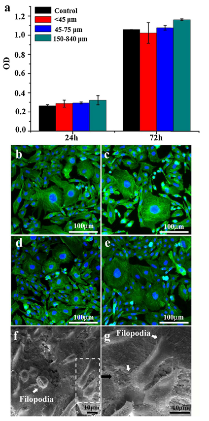
(2)Han Y, Lian M, Sun B, Jia B, Wu Q, Qiao Z, Dai K. Preparation of high precision multilayer scaffolds based on Melt Electro-Writing to repair cartilage injury. Theranostics 2020; 10(22):10214-10230. doi:10.7150/thno.47909.
After 21 days of culturing, the cell-bearing scaffolds (n = 4 per group) were fixed in 4% paraformaldehyde for 2 h. Triton-X (0.1%) was used to soak the scaffolds for 5 min, which were then washed thrice with PBS for 5 min each. Blocking was performed with BSA at room temperature for 1 h. After blotting with water-absorbing paper, primary antibodies (anti-PRG4, anti-CILP, anti-COLII, anti-COLI, and anti-SOX-9) (1:100, Abcam, UK) were added to each sample and incubated at 4 °C overnight. Following washes with PBS, a corresponding secondary antibody (1: 500, Abcam, UK) was added, incubated at room temperature for 1 h, and washed with PBS. The scaffolds were then labeled with FITC phalloidin (1:200, MaoKang, China) and incubated at room temperature for 2 h, followed by incubation with DAPI staining solution (1:1000, MaoKang, China) for 30 min at room temperature. After the final wash with PBS, samples were observed and imaged under a confocal microscope (LEICA, Germany). Analysis and quantification of protein expression was performed using ImageJ.
(3)Li Chengpan et al. On-Chip Replication of Extremely Early-Stage Tumor Behavior. ACS Appl. Mater. Interfaces 2021, 13, 17, 19768–19777 Phalloidin-FITC, and DAPI dyes were purchased from Shanghai Maokang Biotechnology Ltd.
[4]Ming, Ping Deng, et al. “Preparation and Characterization of Silk Fibroin-Based Hybrid Vascular Tissue Engineering Film.” Materials Science Forum, vol. 996, Trans Tech Publications, Ltd., June 2020, pp. 64–69. Crossref, doi:10.4028/www.scientific.net/msf.996.64. FITC Phalloidin was purchased from Shanghai Mao Kang biotechnology Co., Ltd., ( Shanghai, China)
[5]Cheng panLi et al. Alginate core–shell microcapsule reduces the DMSO addition-induced osmotic damage to cells by inhibiting cellular blebs. Chinese Journal of Chemical Engineering. Volume 33, May 2021, Pages 249-255
[6] Xia, C., Ming, P., Zhou, A. et al. Supramolecular self-assembly of oligopeptide hybrid films with liquid crystal texture: effects on cell behaviour for vascular grafts. Bull Mater Sci 44, 197 (2021). https://doi.org/10.1007/s12034-021-02470-x
Each sample was stained with DAPI and FITC-phalloidin reagents (Shanghai Mao Kang Biotechnology Co. Ltd).
[7] Wei Cui, Qibin Song, Huhu Su, Zhiqing Yang, Rui Yang, Na Li, Xing Zhang. Synergistic effects of Mg-substitution and particle size of chicken eggshells on hydrothermal synthesis of biphasic calcium phosphate nanocrystals[J]. Journal of Materials Science & Technology, 2020, 36(0): 27-36 https://doi.org/10.1016/j.jmst.2019.04.038
[8] MC3T3-E1 cells with the initial density of 104 cells/cm2 were placed in culture plates. After culture with BCP extracts using different particle sizes of 45 μm, 45-75 μm, 150-840 μm and without BCP extracts (control group) for 24 h, cells washed with phosphate buffered saline (PBS) were fixed with 4% paraformaldehyde at 4 °C for 15 min. The cells were stained with 10 ug/ml FITC-phalloidin (Mao Kang, Biotechnology, co, LTD, Shanghai, China) for 50 min and counterstained with 100 mg/ml 4′,6-diamidino-2-phenylindole (DAPI, Dingguo Changsheng Biotechnology, co, LTD, Beijing, China) for 15 min at room temperature after washed with PBS for three times. Finally, the fluorescent staining of cells was observed by an Eclipse Ti fluorescence inverted microscope (Nikon Instruments Inc., Tokyo, Japan).
[9]LeiCao et al. Plasma spray of biofunctional (Mg, Sr)-substituted hydroxyapatite coatings for titanium alloy implants. Journal of Materials Science & Technology. Volume 35, Issue 5, May 2019, Pages 719-726
MC3T3-E1 cells were placed in culture plates at an initial density of 1 × 10⁴ cells/cm2. After culture for 48 h, cells were washed with phosphate buffered saline (PBS) three times, and fixed with 4% paraformaldehyde at 4 °C for 15 min. The fixed samples were then washed with PBS three times and stained with 10 μg/ml FITC-phalloidin (Mao Kang, Biotechnology, co, LTD, Shanghai, China) for 50 min at room temperature.
Fig. 7. Immunofluorescence staining of MC3T3-E1 cells cultured with (a, b) normal culture medium and (c, d) (Mg, Sr)-HA coating extract for 48 h. (b, d) show the partially enlarged images from the white dash boxes from (a, c). The cell nuclei and smooth muscle alpha-actin are stained with DAPI (blue) and FITC-phalloidin (green), respectively.
[10]Huo-Liang Zheng, Wen-Ning Xu, Peng-Bo Chen, Lei-Sheng Jiang, Xin-Feng Zheng, Sheng-Dan Jiang, "Increased Expression of Prolyl Endopeptidase Induced by Oxidative Stress in Nucleus Pulposus Cells Aggravates Intervertebral Disc Degeneration", Oxidative Medicine and Cellular Longevity, vol. 2022, Article ID 9731800, 16 pages, 2022. https://doi.org/10.1155/2022/9731800
DAPI (Beyotime) and FITC-conjugated phalloidin (MKBio) were used to stain the nuclei and cytoskeletons. Images were obtained using an Olympus microscope.
|
|||||||||||||||||||||||||||||||||||||||||||||||||||||||||||||||||||||||||||||||||||||