| 目录号 | MX4001-10MG | 售价 | 658.00元 | |||||||||||||||||||||||||||||||||||||||||||||||||||||||||||||||||||||||||||
| 规格 | 10mg | 运输温度 | 冰袋运输 | |||||||||||||||||||||||||||||||||||||||||||||||||||||||||||||||||||||||||||
| 其他名称 | DiO perchlorate; DiOC18(3); 3,3-Dioctadecyloxacarbocyanine perchlorate; | 保存温度 | 2-8ºC避光干燥保存 | |||||||||||||||||||||||||||||||||||||||||||||||||||||||||||||||||||||||||||
| CAS号 | 34215-57-1 | 有效期 | 1年 | |||||||||||||||||||||||||||||||||||||||||||||||||||||||||||||||||||||||||||
| 应用 | 绿色细胞膜探针,神经示踪研究 | 订购数量 |
|
|||||||||||||||||||||||||||||||||||||||||||||||||||||||||||||||||||||||||||
|
产品简介: DiO (DiOC18(3)) 细胞膜绿色荧光探针
搜索关键词: 细胞膜荧光探针,DiI,DiO,DiD,DiR,DiA,CM-DiI,神经示踪,传统细胞膜荧光探针
DiO订购信息:
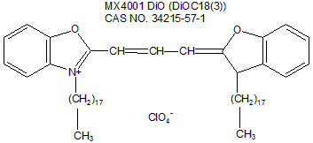
DiO绿色荧光探针基本特性: 1. 化学名:Benzoxazolium, 3-octadecyl-2-[3-(3-octadecyl-2(3H)-benzoxazolylidene)-1-propenyl]-, perchlorate 2. 同义名:DiO perchlorate; DiOC18(3); 3,3-Dioctadecyloxacarbocyanine perchlorate; 3. 推荐滤光器:Omega-XF100, XF23, Chroma-41001, 31001 4. CAS NO:34215-57-1 5. 分子式:C53H85ClN2O6 6. 分子量:881.72g/mol 7. 外观:橙黄至橙色固体 8. 纯度:≥98% 9. Ex/Em:484/501 nm(甲醇) 10. 溶解性:溶于DMF,DMSO,甲醇
11. 化学结构图:
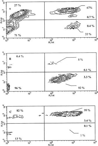
应用示例(DiO,来自文献): 文献一:Piriou L et al. Design of a flow cytometric assay for the determination of natural killer and cytotoxic T-lymphocyte activity in human and in different animal species. Cytometry. 2000 Dec 1;41(4):289-97.
使用方法: For DIOC18(3) labeling, 1 ml of complete medium containing 106 cells was mixed with 10 μl of 3 mM DIOC18(3). The cells were incubated at 37°C for 15 min and rinsed three times with PBS. Cell labeling and cell viability were confirmed by flow cytometry before use in the test. Cell viability was assessed by PI, which permeates only through the membrane of dead cells and emits a red fluorescence (10 μg/ml, optimal concentration determined after a calibration assay. The test was performed when cells were labeled homogeneously and cell viability was higher than 80%.
Fig 1. FCA of target cells in an NK cell cytotoxicity assay. Four cell subpopulations are shown by fluorescent staining with DIOC18(3) (green: x axis, FL1) and PI (red: y‐axis, FL3). The fluorescence contour plot cytogram (FL1 versus FL3) shows target cells labeled with DIOC18(3) (FL1) versus the dead cells labeled with PI (FL3). Quadrant 1 = lysed effector cells; quadrant 2 = live effector cells; quadrant 3 = lysed K562 target cells; quadrant 4 = live K562 target cells.
文献二、Romero-Pérez GA et al. Orally Administered Salacia reticulata Extract Reduces H1N1 Influenza Clinical Symptoms in Murine Lung Tissues Putatively Due to Enhanced Natural Killer Cell Activity. Front Immunol. 2016 Mar 31;7:115.
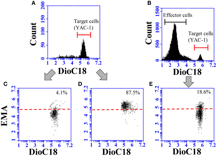
使用方法: YAC-1 cells were cultured in 75-cm2culture flasks containing 10 mL of culture media and incubated in a humidified chamber (37°C, 5% CO2). After 3-day incubation, YAC-1 cells were stained for 15 min at 37°C with 10μM Dioc18 [3,3′-dioctadecyloxacarbocyanine perchlorate] to cause green fluorescence of cell membranes. YAC-1 cells were then washed twice with 1mL HBSS and seeded in 100 μL of culture media in 96-well culture plates in a concentration of 1.0 × 10⁴ cells (for coculture with splenocytes) or 5.0 × 10³cells (for coculture with pulmonary cells) per well.
For the NK activity assays, YAC-1 cells were used as target cells, and splenocytes and pulmonary cells as effector cells. One hundred microliters each of splenocyte and pulmonary cell suspensions were added to each well containing DioC18-labeled YAC-1 cells to achieve 25:1 and 10:1, and 10:1 and 5:1 effector cell:target cell (E:T) ratios, respectively. After incubation for 4 h, 10 μL of ethidium monoazide bromide (EMA, 0.5 ng/mL) solution was added to each well for nuclear staining of dead cells. The ethidium was bound to nuclei by photo affinity using a 26-W fluorescent light (5 cm distance) for 10 min at room temperature. After washing twice with PBS containing 0.5% BSA, the cells were fixed with 2% PFA overnight. Each sample was incubated in duplicate and analyzed using an Accuri C6 flow cytometer.
Fig 2. Representative images of flow-cytometric analysis for the determination of NK activity. To discriminate the target cells, live gate (red line) was set in the FL1 histogram on the green fluorescent (A,B). The target cells were further analyzed with a dot plot of FL1 and FL3 (C–E).
传统细胞膜探针: 1. DiI, DiO, DiD和DiR DiI, DiO, DiD和DiR作为一类长链的亲脂性二烷基碳菁类染料(Dialkylcarbocyanines)荧光染料家族,用于标记细胞膜以及其他脂溶性生物结构,尤其是DiI,广泛用于活体和固定组织及细胞的神经元逆行性和顺行性示踪。
作为一类环境敏感型荧光染料,当它们与膜结合或者与亲脂性生物分子(例如蛋白质,虽然在水中其荧光强度很弱)结合时,其荧光强度显著增强。一旦进入细胞后,它们在细胞内质膜中逐步扩散,最佳浓度条件下可将整个细胞染色。这些染料具很高的淬灭系数,偏光依赖性和很短的激发寿命。
四种染料呈现不同的荧光颜色,DiI(橙色荧光)、DiO(绿色荧光)、DiD(红色荧光)、DiR(深红色荧光),为活细胞多色彩荧光成像分析和流式细胞术提供了一种便捷的工具。DiO和DiI分别用标准FITC和TRITC滤光器检测,可结合使用。DiD可被633 nm氦-氖(He-Ne)激光激发,具有比DiI更长的激发和发射光波长,特别适合用于标记具本底荧光的细胞和组织。而,DiR在体内活体成像或者示踪中意义非凡,因其所发射的红外光可以高效地穿过细胞和组织,并且在红外光范围内,其本底荧光水平很低。
2. DiA DiA为亲脂性氨基苯乙烯基染料(dialkylaminostyryl dye),常用于神经元的示踪,可与DiI联合使用进行神经细胞的双色标记。研究发现,某些情况下当DiO不能标记固定组织时,DiA可得到良好标记效果。DiA与磷脂双层膜结合后,光谱迁移明显加强。正由其发射光谱非常广,在适当的滤光片下,可以绿色、橙色,甚至红色荧光被检测到。
产品订购信息:
细胞膜探针光谱和检测滤片:
— —Written/Edited by V. Shallan【版权归MKBio懋康所有】
上海懋康生物科技有限公司是一家涉足于生命科学和生物技术领域研究的试剂、仪器和实验室消耗品与实验服务工作,主要从事细胞生物学、植物学、分子生物学、免疫学、生物化学、蛋白组学。生物制药与诊断试剂研发生产等领域。 本公司秉承“以人为本,以诚为信、合同守信”的经营理念。坚持"品质保障"的原则为广大客户提供优质产品。
|
||||||||||||||||||||||||||||||||||||||||||||||||||||||||||||||||||||||||||||||