| 目录号 | MX4002-10MG | 售价 | 348.00元 | |||||||||||||||||||||||||||||||||||||||||||||||
| 规格 | 10mg | 运输温度 | 冰袋运输 | |||||||||||||||||||||||||||||||||||||||||||||||
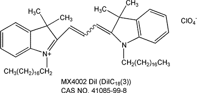
| 其他名称 | DiI perchlorate; DiIC18(3); 1,1′-Dioctadecyl-3,3,3′,3′-tetramethylindocarbocyanine perchlorate; | 保存温度 | 4ºC干燥避光保存 | |||||||||||||||||||||||||||||||||||||||||||||||
| CAS号 | 41085-99-8 | 有效期 | 一年 | |||||||||||||||||||||||||||||||||||||||||||||||
| 应用 | 细胞膜红色探针 | 订购数量 |
|
|||||||||||||||||||||||||||||||||||||||||||||||
|
产品简介: DiI (DiIC18(3)) 细胞膜橙红色荧光探针(神经元示踪研究)
温馨提示:见细胞膜荧光探针专题,选择你想要的最适膜标记探针。 关键词:细胞膜荧光探针,DiI,DiO,DiD,DiR,DiA,CM-DiI,神经示踪,传统细胞膜荧光探针 订购信息:
产品描述: DiI, DiO, DiD和DiR作为一类长链的亲脂性二烷基碳菁类染料(Dialkylcarbocyanines)荧光染料家族,用于标记细胞膜以及其他脂溶性生物结构。作为一类环境敏感型荧光染料,当它们与膜结合或者与亲脂性生物分子(例如蛋白质,虽然在水中其荧光强度很弱)结合时,其荧光强度显著增强。一旦进入细胞后,它们在细胞内质膜中逐步扩散,于最佳浓度条件下可将整个细胞均匀染色。这些染料具很高的淬灭系数,偏光依赖性和很短的激发寿命。 四种染料呈现不同的荧光颜色,DiI(橙色荧光)、DiO(绿色荧光)、DiD(红色荧光)、DiR(深红色荧光),为活细胞多色彩荧光成像分析和流式细胞术提供了一种便捷的工具。DiO和DiI分别用标准FITC和TRITC滤光器检测,可结合使用。其中,DiI及其衍生物由于极其低的细胞毒性,应用最为广泛。不仅普遍用于活体和固定组织及细胞的神经元逆行性和顺行性示踪分析,还可以用于检测细胞的融合和粘附,检测发育或移植过程的细胞迁移,通过FRAP(荧光光漂白恢复技术)检测脂质在细胞膜上的扩散过程,检测细胞毒性,以及标记脂蛋白如LDL和HDL等。 本品以DiI的高氯酸盐形式提供,英文全名:1,1′-Dioctadecyl-3,3,3′,3′-tetramethylindocarbocyanine perchlorate,CAS NO.41085-99-8,纯度≥95%(TLC),适用于荧光检测研究。
DiI橙红色荧光探针基本特性: 1) 同义名:DiI perchlorate; DiIC18(3);1,1′-Dioctadecyl-3,3,3′,3′-tetramethylindocarbocyanine perchlorate; 2) CAS NO:41085-99-8 3) 分子式:C59H97ClN2O4 4) 分子量:933.87 g/mol
5)纯度:≥95%(TLC)
保存与运输方法 保存:4ºC干燥避光保存,有效期一年。 运输:冰袋运输。 注意事项 1)荧光染料均存在淬灭问题,请尽量注意避光,以减缓荧光淬灭。 2)为了您的安全和健康,请穿实验服并戴一次性手套操作。 使用方法 【注意】以下使用方法仅用作参考,可根据具体的实验条件做出调整。 1.染色液制备 1)储存液制备:用DMSO或乙醇配置浓度1~5 mM的储存液。例如,取5mg DiI(Mw:933.87 g/mol)溶于1.07ml无水DMSO中,充分溶解,即得到5mM的储存液。【注意】未使用的储存液分装储存在-20℃,避免反复冻融。 2)工作液制备:用合适的缓冲液(如:无血清培养基,HBSS或PBS)稀释储存液,调整到1~5 μM的工作浓度。【注意】工作液最终浓度需要根据不同细胞系和实验体系来优化。建议从推荐浓度开始,以10倍范围为区间进行最优浓度的摸索。 2.悬浮细胞染色 1)加入适当体积的染色工作液重悬细胞,使其密度为1×10⁶/mL。 2)37℃孵育细胞2~20min,不同的细胞最佳培养时间不同。可以20min作为起始孵育时间,之后优化以保证得到均一化的标记结果。 3)孵育结束,按1000~1500 rpm离心5min。 4)去除上清液,之后轻柔加入37℃预热的生长培养液重悬细胞。 5)再重复3),4)步骤两次。 3.贴壁细胞染色 1)将贴壁细胞培养于无菌盖玻片上。 2)从培养基中移走盖玻片,滤掉过量培养液,将盖玻片放在潮湿的小室内。 3)在盖玻片的一角加入100μL的染色工作液,轻轻晃动使染料均匀覆盖所有细胞。 4)37℃孵育细胞2~20min,不同的细胞最佳培养时间不同。可以20min作为起始孵育时间,之后优化以保证得到均一化的标记结果。 5)吸掉染色工作液,用培养液洗盖玻片2~3次,每次用预温的培养基覆盖所有细胞,孵育5~10min,然后吸走培养基。 4.显微镜检测 1)DiD,DiO,DiI,DiR和DiS滤光器的选择参见下表:
2)多色染料的同时检测,滤光器按照以下设定: a) DiI和DiO=Omega XF52,Chroma 51004; b) DiI和DiD=Omega XF92,Chroma 51007; c) DiI,DiO和DiD=Omega XF93,Chroma 61005; 5.流式细胞仪检测 经DiO,DiI,DiD和DiR染色的细胞分别用流式细胞仪的FL1,FL2,FL3或FL4通道检测。
应用示例(来自文献): 文献一、Wexler-Cohen Y et al. Membrane-Anchored HIV-1 N-Heptad Repeat Peptides Are Highly Potent Cell Fusion Inhibitors via an Altered Mode of Action. PLoS Pathog. 2009 Jul;5(7):e1000509.
使用方法(Cell-Cell Fusion Inhibition Assay): Jurkat E6-1 and Jurkat HXBc2 cells were labeled with DiI and DiD lipophilic fluorescent probes, respectively. The two cell populations were co-incubated, in a ratio of 1∶1, for 6 h in the presence of eight different concentrations of the inhibitory peptides.Prior to measurements the cells were washed, spinned, dissolved in PBS, and put on ice. Cells co-incubated without the presence of peptides served as an optimal fusion reference. Unlabeled cells that were handled similarly served as an intrinsic fluorescence control.
Cells labeled separately with DiI or DiD were used to adjust the optimal separation of fluorescent signals. Jurkat HXBc2 cells labeled with DiI were co-incubated with Jurkat HXBc2 cells labeled with DiD for a fusion background that was subtracted from the measurements of the experiment.The following alterations were applied to the original protocol: (i) 5 µL of a 1 mg/mL DiI or DiD solution in dimethylsulfoxide (DMSO) was added to 1 mL of 4×10⁶ cells/mL Jurkat E6-1 or Jurkat HXBc2 cells, respectively.(ii) For each data point 150,000 events were collected. Measurements were performed on a FACSort machine, upgraded to a FACSCalibur cell analyzer (Becton Dickinson).
文献二、Mazzoni M et al. Distribution, organization and innervation of gastric MALT in conventional piglet. J Anat. 2011 Nov;219(5):611-21.
使用方法(Tissue preparation and DiI tracing in fixed tissue): Small crystals of DiI were diluted at 3% in 100% ethanol and evaporated onto small glass beads (about 200 μm). The glass beads were placed in the middle of the gastric folliclesto detect if neurones projecting to the follicles were present. In other samples, after gently removing the overlying epithelium by the use of entomological forceps, glass beads were inserted into the lamina propria to identify whether neurones projecting to the mucosal layers were present.
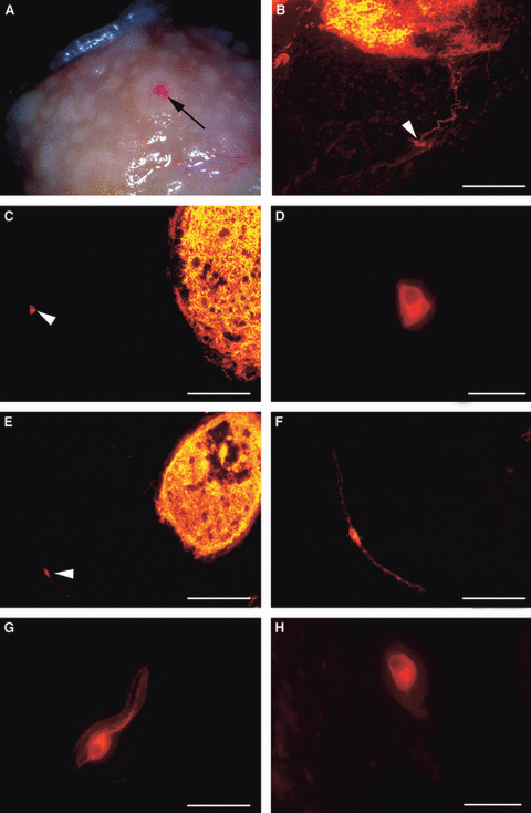
Fig 1. Tangential sections of pig gastric mucosa at the level of the diverticulum.(A) Stereomicroscopy showing DiI‐coated beads (arrow) applied to a follicle in fixed tissue after 8 months of incubation. (B,C,E) DiI crystals applied to the follicles; note that the tracer was homogeneously distributed inside the follicles. (B) Labelled neurones (arrowhead) and processes entering the labelled follicle. (C) Polyhedral‐labelled neurone (arrowhead) more than 400 μm from a labelled follicle. (D) Higher magnification of the neurone shown in (C). (E) Elongated neurone (about 400 μm from the labelled follicle) along a thin nervous fascicle (arrowhead). (F) Higher magnification of the neurone shown in (E). (G,H) Two labelled neurones with an ovoid shape. Note that the neurones showed smoothly contoured soma and an eccentrically located nucleus (D,G,H,)
— —Written/Edited by V. Shallan【版权归MKBio懋康所有】
上海懋康生物科技有限公司是一家涉足于生命科学和生物技术领域研究的试剂、仪器和实验室消耗品与实验服务工作,主要从事细胞生物学、植物学、分子生物学、免疫学、生物化学、蛋白组学。生物制药与诊断试剂研发生产等领域。 本公司秉承“以人为本,以诚为信、合同守信”的经营理念。坚持"品质保障"的原则为广大客户提供优质产品。
|
||||||||||||||||||||||||||||||||||||||||||||||||||