

| 目录号 | MX4558-50UG | 售价 | 1300元 | ||||||||||||||||||||||||||||||||||||||
| 规格 | 50μg | 运输温度 | 冰袋运输 | ||||||||||||||||||||||||||||||||||||||
| 其他名称 | RhoNox-1 | 保存温度 | -20℃避光干燥保存 | ||||||||||||||||||||||||||||||||||||||
| CAS号 | N/A | 有效期 | 至少1年 | ||||||||||||||||||||||||||||||||||||||
| 应用 | 亚铁离子探针「倾向定位高尔基体」 | ||||||||||||||||||||||||||||||||||||||||
|
产品简介:
FeRhoNox-1 (Fe2+indicator) 亚铁离子荧光探针
重要提醒(购买或初次使用前)请务必查阅:
一、荧光染料(粉末形式,特别是对氧敏感探针)配制储存液的注意事项
1)荧光探针在固体(粉末)状态很稳定,按照说明书要求温度来保存,效期内使用即可。
2)荧光探针用有机溶剂比如:DMSO,溶解配制成储存液之后,一般来说,放到-20℃保存(2-3个月内使用,甚至更短,具体可咨询);放到-80℃保存(6个月内使用,具体可咨询)。前提是,使用的有机溶剂必须是高质量且无水的,特别是DMSO,必须是新鲜开封。
3)配制的储存液请务必用密封性好且螺旋盖的低容量冻存管保存(不可用EP管),至少按照5-10ul/管来分装,避光冻存。
4)对于某些特殊化合物(对空气敏感或存在不稳定结构),可能保存周期特别短,甚至只能当天使用。这些化合物说明书上会有说明。
有更多信息,请联系我司工作人员来核实。
二、荧光染料(以溶于有机溶剂的储存液形式提供)的注意事项
1) 以溶于有机溶剂的储存液形式的荧光探针相对来说是比较稳定的化合物;但收到这类产品,也需用户根据单次用量(5-10ul/管来分装),-20℃以下密封避光保存,减少反复冻融次数。
2) 请务必用密封性好且螺旋盖的低容量冻存管保存(不可用EP管)。务必避光。
有更多信息,请联系我司工作人员来核实。
产品标签 FeRhoNox-1;FerroOrange;Labile ferrous ion 不稳定铁(II)离子;Fe2+荧光探针;Ferroptosis 铁死亡;C11 BODIPY 581/591 脂质过氧化探针;
产品信息
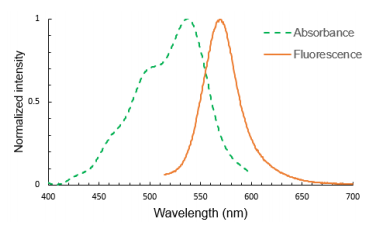
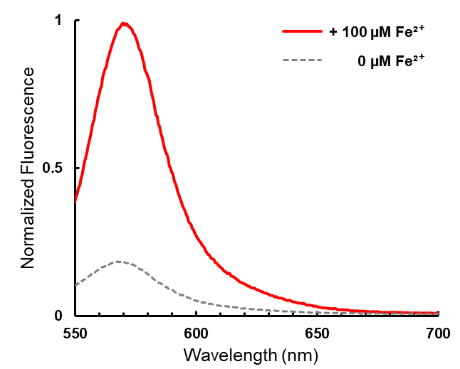
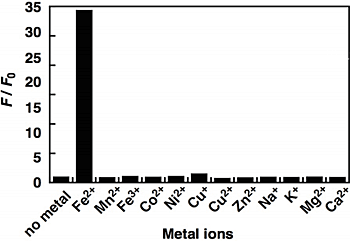
产品描述 FeRhoNox-1,也称为RhoNox-1,是一种活跃的荧光探针,特异性检测不稳定的铁(II)离子(Fe2+)。一旦与Fe2+反应后,不可逆的生成一种橙色(红色)荧光产物(Absmax=540nm,FLmax=575nm,图1.FeRhoNox-1的光谱特征)。生理浓度下的铁(III)离子(Fe3+)或其它除铁离子以外的二价金属离子都不会使其荧光增强(见图2FeRhoNox-1的选择性).FeRhoNox-1的反应特异性)。FeRhoNox-1具细胞膜渗透性和高选择性,适用于活细胞内Fe2+的检测,倾向定位在高尔基体。
图1. FeRhoNox-1的光谱特征。FeRhoNox-1与Fe2+反应后吸收和发射光谱(上)。FeRhoNox-1在37℃,与Fe2+反应1h后,荧光明显增强。最大荧光峰约在575nm。
图2. FeRhoNox-1的选择性。FeRhoNox-1仅与Fe2+反应。
保存与运输方法 保存:-20℃避光干燥保存,至少1年有效。 运输:冰袋运输。
注意事项
使用说明 1. 需要自行准备的材料 1.1细胞培养级或超纯DMSO(比如:MS4601A-100ML)【强烈建议将高纯的DMSO分装成单次用量保存在极低温冷冻室内,比如-80℃,避免吸潮。降解的DMSO可能会增加FeRhoNox-1的背景信号】 1.2合适的清洗和观察缓冲液(比如:PBS, pH 7.4;HBSS;等)。不要含酚红。
2. 探针准备 2.1从冰箱取出FeRhoNox-1,置于室温回温至少30min,将其置于微量离心机内低速离心。将瓶内的粉末离心到管底后,再开盖。 2.2往一管FeRhoNox-1(50μg)内加入109μl高质量DMSO,用枪反复吹吸5次或以上,使其完全溶解即得到1mMFeRhoNox-1储存液。建议单次用完储存液,若实在用不完,请根据单次用量分装,置于-80℃避光保存。用中性缓冲液来稀释储存液。 2.3于正式实验前,用HBSS或其他中性缓冲液来稀释1mMFeRhoNox-1储存液到所需工作浓度(比如:5μM),工作液需现配现用,尽快用完。【注意:酸性溶液会氧化FeRhoNox-1,严重影响探针的效率】。
3. 染色步骤 4. 荧光检测 对于荧光激发:通用的绿色激发滤片比如Cy3或四甲基罗丹明(TMR)检测用的滤片。 对于激光激发:532nm或543nm激光器比较适合。发射波长为570nm左右。
相关产品
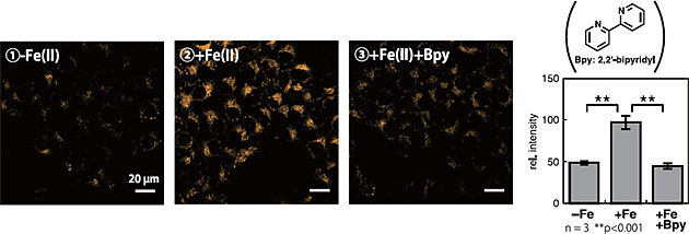
附录F eRhoNox-1的染色示例 I. HepG2细胞
图3. FeRhoNox-1在HepG2活细胞内的成像检测。①-Fe(II):细胞未添加亚铁离子(100μM硫酸亚铁铵);②+Fe(II):细胞加载亚铁离子;③+Fe(II)+Bpy:细胞加载亚铁离子,之后用加入铁离子螯合剂Bpy。图②荧光增强,而图③荧光降低。此结果与FeRhoNox-1特异性检测Fe(II)的特征一致。
— —Written/Edited by V. Shallan【版权归MKBio懋康所有】
上海懋康生物科技有限公司是一家涉足于生命科学和生物技术领域研究的试剂、仪器和实验室消耗品与实验服务工作,主要从事细胞生物学、植物学、分子生物学、免疫学、生物化学、蛋白组学。生物制药与诊断试剂研发生产等领域。 本公司秉承“以人为本,以诚为信、合同守信”的经营理念。坚持"品质保障"的原则为广大客户提供优质产品。
引用文献:
[1] Lu Zhou, Peng Yu, Ting-ting Wang, Yi-wei Du, Yang Chen, Zhen Li, Man-lin He, Lan Feng, Hui-rong Li, Xiao Han, Heng Ma, Hong-bao Liu, "Polydatin Attenuates Cisplatin-Induced Acute Kidney Injury by Inhibiting Ferroptosis", Oxidative Medicine and Cellular Longevity, vol. 2022, Article ID 9947191, 14 pages, 2022. https://doi.org/10.1155/2022/9947191
Method: FeRhoNox-1 fluorescent probe (MX4558) was purchased from Maokang Biotech (Shanghai, China).FeRhoNox-1,which is a turn-on fluorescent probe specific for the detection of labile iron Fe2+, was used to detect intracellular LIP, and the cellular distribution of FeRhoNox-1 was consistent with Golgi [41]. HK-2 cells were grown to confluence in 35 mm laser confocal petri dishes in DMEM, and PD (40 μM) or Fer-1 (1 μM) was added in the absence or presence of cisplatin (20 μM). Cells were incubated with 5 μM FeRhoNox-1 for 1 h prior to assays. Cells were washed twice with PBS before staining nuclei with Hoechst 33342. The fluorescence was immediately observed with a confocal laser-scanning microscope (CLSM, ECLIPSE Ti, Nikon, Tokyo, Japan).
[2] Jin R, Yang R, Cui C, Zhang H, Cai J, Geng B, Chen Z. Ferroptosis due to Cystathionine γ Lyase/Hydrogen Sulfide Downregulation Under High Hydrostatic Pressure Exacerbates VSMC Dysfunction. Front Cell Dev Biol. 2022 Feb 3;10:829316. doi:
10.3389/fcell.2022.829316. PMID: 35186934; PMCID: PMC8850391. Method: For FeRhoNox-1 staining, 5 μM of the FeRhoNox-1 staining working solution (MX4558 and MKBIO)was added and incubated in a 37°C, 5% CO2 incubator for 60 min after treatment for 24 h in a hydrostatic pressure chamber. After washing three times with PBS, cells were imaged with a confocal microscope.
[3] Zhang X, Ma Y, Ma J, Yang L, Song Q, Wang H, Lv G. Glutathione Peroxidase 4 as a Therapeutic Target for Anti-Colorectal Cancer Drug-Tolerant Persister Cells. Front Oncol. 2022 Jun 3;12:913669. doi: 10.3389/fonc.2022.913669. PMID: 35719967; PMCID: PMC9203854.
Method: FeRhoNox-1 (an Fe2+ indicator, Cat# MX4558) was purchased from MKBio (Shanghai, China). Ferrous Iron Staining
Cells were incubated for 1 h at 37°C with FeRhoNox-1 (an Fe2+ indicator) to detect ferrous iron. The cells were then harvested by trypsinization, and the level of ferrous iron was determined by imaging using a confocal microscope or by flow cytometry analysis.
[4] Fang J, Yuan Q, Du Z, Fei M, Zhang Q, Yang L, Wang M, Yang W, Yu J, Wu G, Hu J. Ferroptosis in brain microvascular endothelial cells mediates blood-brain barrier disruption after traumatic brain injury. Biochem Biophys Res Commun. 2022 Sep 3;619:34-41. doi: 10.1016/j.bbrc.2022.06.040. Epub 2022 Jun 14. PMID: 35728282. Method: Intracellular Fe 2+ levels were evaluated by using FeRhoNox-1 probe (MKBio, Shanghai). 24 h after SI or RSL3 incubation, culture medium was washed by PBS and exchanged for HBSS
Lai Y, Zeng F, Chen Z, et al. Shikonin Could Be Used to Treat Tubal Pregnancy via Enhancing Ferroptosis Sensitivity. Drug Design, Development and Therapy. 2022 ;16:2083-2099. DOI: 10.2147/dddt.s364441. PMID: 35800255; PMCID: PMC9255906.
Method: Labile Iron Pool (LIP) Assay “Labile iron” (which is primarily in the ferrous (Fe2+) form) is a small, transitional pool of intracellular iron, and commonly termed “LIP”. LIP release was measured using a FeRhoNox-1™ (Fe2+ indicator) fluorescent probe (MKBio, Beijing, China). HTR-8/SVneo cells were plated in six-well plates, loaded with FeRhoNox-1 (5 μM) for 30 min at 37°C and then washed thrice with Hanks’ balanced salt solution. Cells were observed under a fluorescence microscope (Olympus).
[5] Cui J, Zhou Q, Yu M, Liu Y, Teng X, Gu X. 4-tert-butylphenol triggers common carp hepatocytes ferroptosis via oxidative stress, iron overload, SLC7A11/GSH/GPX4 axis, and ATF4/HSPA5/GPX4 axis. Ecotoxicol Environ Saf. 2022 Sep 1;242:113944. doi: 10.1016/j.ecoenv.2022.113944. Epub 2022 Aug 1. PMID: 35926411.
[6] Method: Intracellular Fe2+ determinationFeRhoNox-1 (Fe2+ Indicator) fluorescent probe (Maokang Biotechnology Co., Ltd., Shanghai, China) was used to detect intracellular Fe2+ content. The cells inoculated in cell culture dishes were treated separately (Please see 2.9 for details), and then were washed twice with PBS. The FeRhoNox-1 (5 μM) was added in the medium and the cells were incubated for 60 min. Finally, images were obtained under the fluorescence microscope, and Image J version 1.43 u software was used to quantify the fluorescence intensity.
{7]Hong H, Lin X, Xu Y, Tong T, Zhang J, He H, Yang L, Lu Y, Zhou Z. Cadmium induces ferroptosis mediated inflammation by activating Gpx4/Ager/p65 axis in pancreatic β-cells. Sci Total Environ. 2022 Nov 25;849:157819. doi: 10.1016/j.scitotenv.2022.157819. Epub 2022 Aug 2. PMID: 35931150.
Method: For cellular Fe 2+ detection, cells were stained with 5 μmol/L FeRhoNox-1 (Fe 2+ indicator, Maokangbio, China) for 60 min at incubator.
[8] Hu Q, Zuo T, Deng L, Chen S, Yu W, Liu S, Liu J, Wang X, Fan X, Dong Z. β-Caryophyllene suppresses ferroptosis induced by cerebral ischemia reperfusion via activation of the NRF2/HO-1 signaling pathway in MCAO/R rats. Phytomedicine. 2022 Jul 20;102:154112. doi: 10.1016/j.phymed.2022.154112. Epub 2022 Apr 22. PMID: 35550220.
Method:The iron level in astrocytes was detected through FeRhoNox-1 (Fe 2+ indicator) (MX4588, Maokangbio, China)
[9] Wu K, Zhang W, Chen H, Wu J, Wang X, Yang X, Liang XJ, Zhang J, Liu D. An Iron Oxyhydroxide-based Nanosystem Sensitizes Ferroptosis by a "Three-pronged" Strategy in Breast Cancer Stem Cells. Acta Biomater. 2023 Feb 21:S1742-7061(23)00084-3. doi: 10.1016/j.actbio.2023.02.015. Epub ahead of print. PMID: 36822484.
FeRhoNox-1 was purchased from MKBio (Shanghai, China).
[10] Weimin Zeng, Zhiru Liu, Charles Amanze, Jinju Cheng, Wanqing Liao, Xueling Wu, Guanzhou Qiu, Qiankun Wang, Zengling Wu, Laichang Zou, Li Shen, In situ detection of Cu2+, Fe3+ and Fe2+ ionsat the microbe-mineral interface during bioleaching of chalcopyrite by moderate thermophiles, Minerals Engineering, Volume 191,2023,107936, ISSN 0892-6875,https://doi.org/10.1016/j.mineng.2022.107936. For Fe 2+ staining, the FeRhoNox-1 Fe 2+ indicator (MKbio, Shanghai, China) was used for single metal labeling to avoid overlap of the emission spectra of the individual dyes.
[11] Pei Z, Qin Y, Fu X, Yang F, Huo F, Liang X, Wang S, Cui H, Lin P, Zhou G, Yan J, Wu J, Chen ZN, Zhu P. Inhibition of ferroptosis and iron accumulation alleviates pulmonary fibrosis in a bleomycin model. Redox Biol. 2022 Oct 18;57:102509. doi: 10.1016/j.redox.2022.102509. Epub ahead of print. PMID: 36302319; PMCID: PMC9614651. The treated cells were collected by trypsin digestion. After the cells were washed with PBS, 5 μM FeRhoNox-1 (MKbio, MX4558) was added to cell suspension, and the cells were incubated at 37 °C for 1 h away from light. The FeRhoNox-1 probe entering the cell would react with Fe2+ to produce an irreversible orange-red fluorescent substance (Ab = 540 nm, FL = 575 nm). After the cells were washed with PBS again, flow cytometry (Fortessa, BD Biosciences) was able to analyze fluorescence in single cell, and the fluorescence intensity was proportional to the concentration of Fe2+ in the cells. [12] Zhang, W.; Lu, J.; Wang, Y.; Sun, P.; Gao, T.; Xu, N.; Zhang, Y.; Xie, W. Canagliflozin Attenuates Lipotoxicity in Cardiomyocytes by Inhibiting Inflammation and Ferroptosis through Activating AMPK Pathway. Int. J. Mol. Sci. 2023, 24, 858. https://doi.org/10.3390/ijms24010858 4.6. Intracellular Ferrous Ion (Fe2+) Measurement Intracellular Fe2+ was detected by a FeRhoNox-1 (MX4558, MKBio, Shanghai, China) fluorescent probe according to the manufacturer’s instructions. After one rinse with PBS buffer, the cells were incubated with 5 µM FeRhoNox-1 for 1 h in an incubator of 5% CO2 at 37 °C. Then, the fluorescence of Fe2+ under a Cy3 excitation filter was observed by a fluorescence microscope after two rinses with PBS buffer. Finally, ImageJ software was used to measure the fluorescence intensity of Fe2+.
[13] Fang, F., Wang, S., Song, Y. et al. Continuous Spatiotemporal Therapy of A Full-API Nanodrug via Multi-Step Tandem Endogenous Biosynthesis. Nat Commun 14, 1660 (2023). https://doi.org/10.1038/s41467-023-37315-0
FeRhoNox-1 (Fe2+ indicator) and hydroxyphenyl fluorescein (HPF) were purchased from Shanghai Maokang Biotechnology Co., Ltd.
|
|||||||||||||||||||||||||||||||||||||||||
 沪公网安备31011202021859号